


![]()
We investigate motion and gait analysis of musculoskeletal disorders and traumas, such as osteoarthritis of the knee, injury of the anterior cruciate ligament (ACL). We also have a strong connection with Clinical Anatomy Laboratory of Department of Anatomy, and have number of in-vitro cadaver studies regarding musculoskeletal disease and trauma. Our group consists of 15 orthopaedic senior residents and 4 graduate students, who are working to complete their PhD degrees.
Takeo Nagura, MD
| Professor | |
|---|---|
| Graduate Year | 1992 |
| Study in Abroad | Stanford University (USA) |
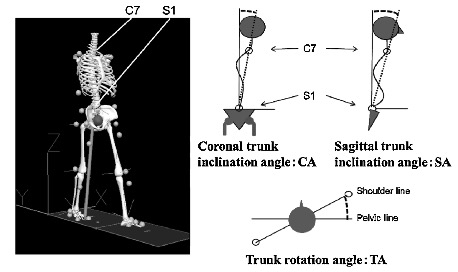
Analysis of 3D Knee Kinematics by Point Cluster Technique
Using cluster markers on the thigh and shank, three-dimensional knee kinematics, including rotation of the tibia relative to the femur, are measured by point cluster technique (PCT). The PCT enables us to evaluate 3D knee kinematics during athletic motion and rehabilitation tasks, that are not possible by fluoroscopic analysis. The PCT is an effective tool of clinical evaluation of the patients following the knee surgery, such as the ACL reconstruction.
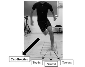
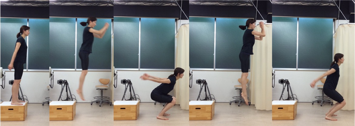
Lower limb mechanics during sports related motion
We use motion capture system to analyze lower limb mechanics during various sports related motions. These data can be used to improve sports performance, prevent lower limb injury, and finally improve treatment of the sports related injuries.
Gait analysis of lower limb disorders
Gait analysis is a powerful tool to evaluate patient’s dynamic function during activities of daily living. Joint moments and kinematics are important index to quantitatively measure the patient’s condition. Knee adduction moment (KAM) during gait is identified as gold standard index for knee osteoarthritis with varus deformity.
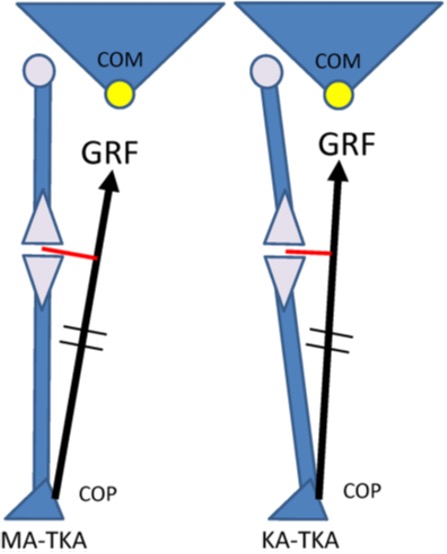
Finite Element Analysis of musculoskeletal disorders
We construct bone models from CT or MRI, and apply them to analyze joint contact force and stress distribution using finite element analysis (FEA). This approach enables us to analyze pathogenesis and onset of various disorders and trauma.
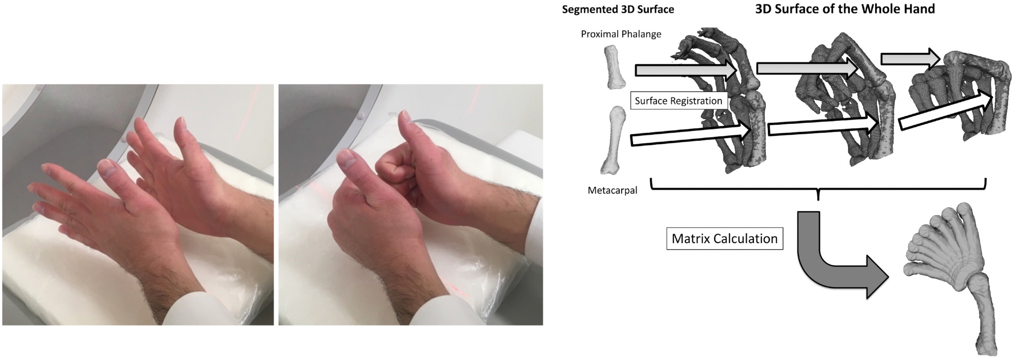
Simulation of the intrinsic muscles of the hand
We developed a simulator to control multiple tendons of the hand, and analyze the function of the intrinsic muscles of the hand. With the traction of the intrinsic muscles, motion area of the thumb was significantly increased compared to the condition without the traction.
Tanikawa H, Matsumoto H, Harato K, Kiriyama Y, Suda Y, Toyama Y, Nagura T. Female recreational athletes demonstrate different knee biomechanics from male counterparts during jumping rope and turning activities. Journal of Orthopaedic Science, 19:104-111, 2014. (0.96)
Kiriyama Y, Wananabe K, Matsumoto M, Toyama Y, Nagura T. Quantification of the spatial strain distribution of scoliosis using a thin-plate spline method. Journal of Biomechanics, 47(1): 302-307, 2014. (2.716)
Kiriyama Y, Matsumoto H, Toyama Y, Nagura T. A miniature tension sensor to measure surgical suture tension of deformable musculoskeletal tissues during joint motion" Journal of Engineering in Medicine (Proceedings of the Institution of Mechanical Engineers, Part H) Vol. 228(2), : 140-148, 2014. (.519)
Takeda K, Hasegawa T, Kiriyama Y, Matsumoto H, Otani T, Toyama Y, Nagura T. Kinematic motion of the anterior cruciate ligament deficient knee during functionally high and low demanding tasks. Journal of Biomechanics, 47: 2526-2530, 2014. (2.716)
Matsumura N, Ogawa K, Kobayashi S, Oki S, Watanabe A, Ikegami H, Toyama Y. Morphological features of humeral head and glenoid version in the normal glenohumeral joint. J Shoulder Elbow Surg. 23, 11, 1724-30, 2014. (2.365)
Tanikawa H, Matsumoto H, Komiyama I, Kiriyama Y, Toyama Y, Nagura T. Comparison of Knee Mechanics among Risky Athletic Motions for Non-contact Anterior Cruciate Ligament Injury. J Applied Biomechanics. epub, 2013 Feb. (1.259)
Harato K, Nagura T, Matsumoto H, Otani T, Toyama Y, Suda Y. Asymmetry of the knee extension deficit in standing affects weight-bearing distribution in patients with bilateral end-stage knee osteoarthritis. Knee Surg Sports Traumatol Arthrosc. epub, 2013 Feb 9. (2.676)
Oki S, Matsumura N, Iwamoto W, Ikegami H, Kiriyama Y, Nakamura T, Toyama Y, Nagura T. Acromioclavicular Joint Ligamentous System Contributing to Clavicular Strut Function - A Cadaveric Study -. J Shoulder Elbow Surg. epub, 2013. (2.747)
Oki S, Matsumura N, Iwamoto W, Ikegami H, Kiriyama Y, Nakamura T, Toyama Y, Nagura T. The function of the acromioclavicular and coracoclavicular ligaments in shoulder motion: a whole-cadaver study. Am J Sports Med. 40, 11, 2617-26, 2012 Nov. (3.792 )
Matsumura N, Nakamichi N, Ikegami H, Nagura T, Imanishi N, Aiso S, Toyama Y. The function of the clavicle on scapular motion: a cadaveric study. J Shoulder Elbow Surg. epub, 2012 May 18. (2.314)
Kuroyanagi Y, Nagura T, Kiriyama Y, Matsumoto H, Otani T, Toyama Y, Suda Y. A quantitative assessment of varus thrust in patients with medial knee osteoarthritis. Knee. 19, 2, 130-4, 2012 Mar. (1.403)
Kokubo T, Hashimoto T, Nagura T, Nakamura T, Suda Y, Matsumoto H, Toyama Y. Effect of the posterior tibial and peroneal longus on the mechanical properties of the foot arch. Foot Ankle Int. 33, 4, 320-5, 2012 Apr. (1.092)
